Gene Cloning Pipeline: Construct Design & In Silico Verification
Introduction
Gene cloning is an essential recombinant DNA technique designed to isolate and generate high-fidelity copies of a specific gene of interest (GOI). The classical procedure involves excising the target sequence using sequence-specific endonucleases, ligating it into a standardized plasmid cloning vector, and introducing the recombinant molecule into an appropriate host strain (such as Escherichia coli DH5
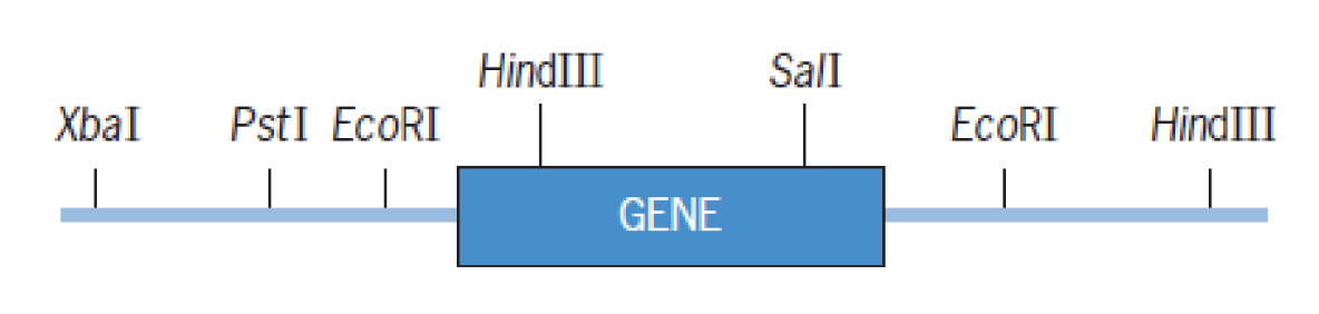
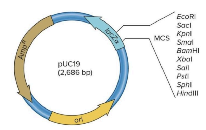
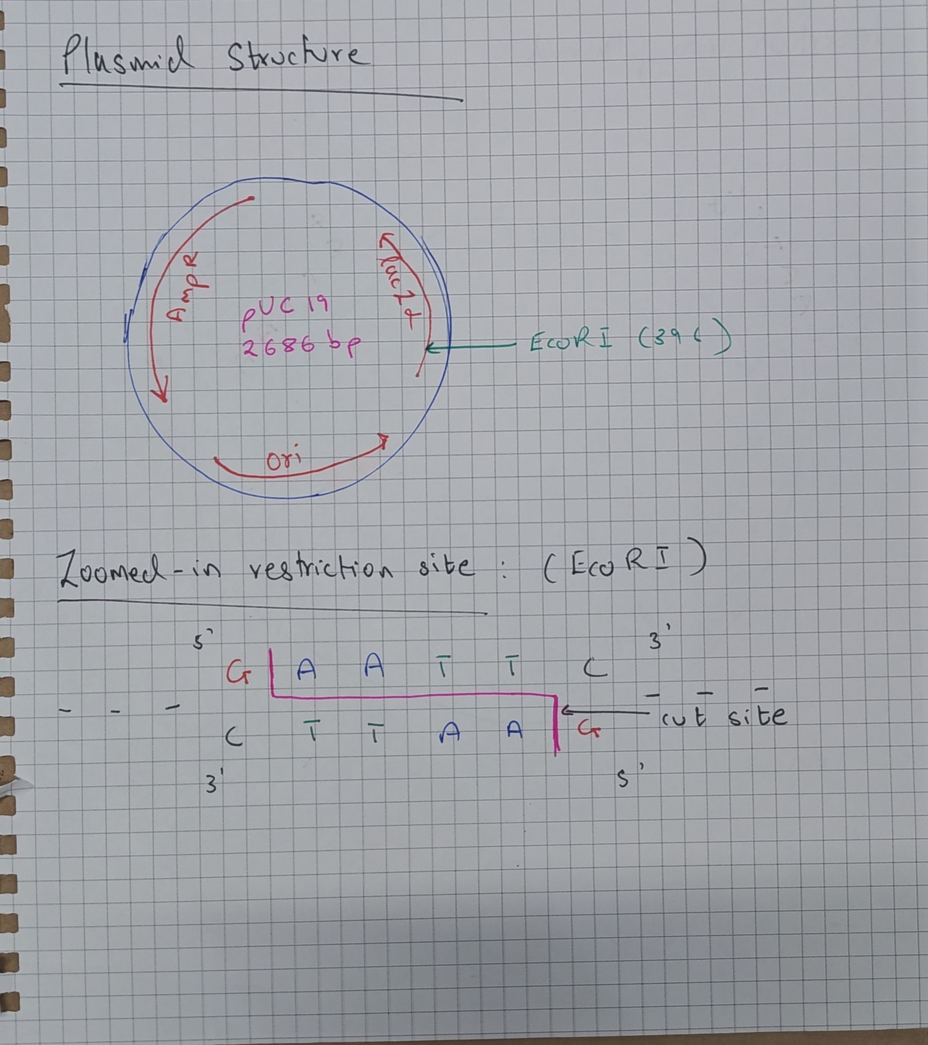
Below are the restriction maps for the target Gene of Interest and the corresponding plasmid cloning vector:
 |  |
|---|---|
| Figure 1: Restriction map of the Gene of Interest flanked by endonuclease cleavage sites. | Figure 2: Structure of the pUC19 cloning vector showing origin of replication (ori), ampicillin resistance ( |
Protocol & Experimental Workflow
Restriction Endonuclease Selection:
The restriction endonuclease EcoRI is selected to excise the GOI. Flanking EcoRI recognition sequences () bracket the complete coding sequence without internal cleavage. Alternative restriction sites either bisect the coding sequence or appear on only one flank. Vector Architecture (pUC19):
The pUC19 vector features an Origin of Replication (ori) and two selectable markers:- Ampicillin resistance (
, -lactamase) -galactosidase -peptide ( ), hosting the Multiple Cloning Site (MCS) including a unique EcoRI restriction site. Insertion of the GOI into the MCS results in insertional inactivation of .
- Ampicillin resistance (
Digestion & Preparative Gel Electrophoresis:
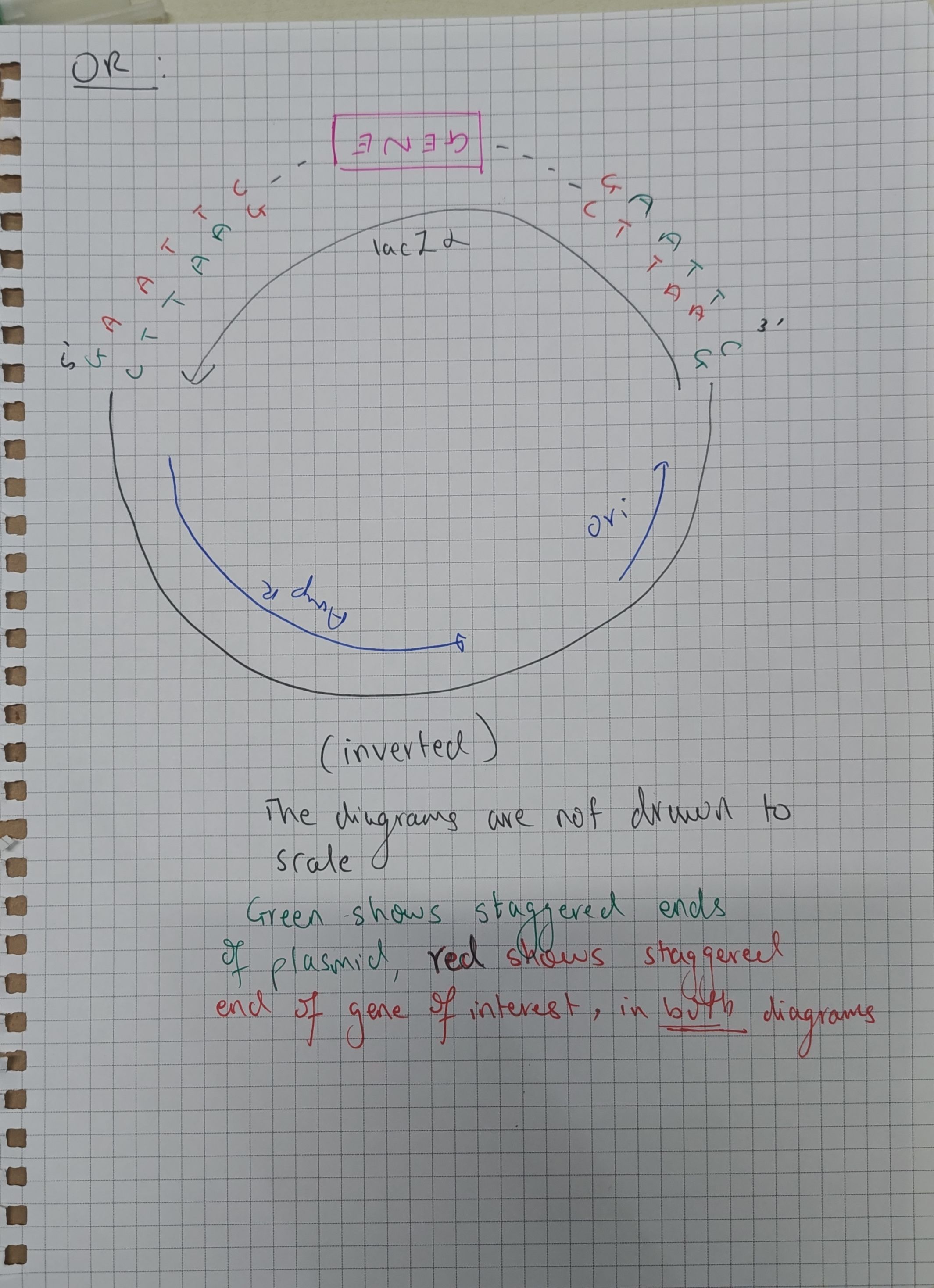
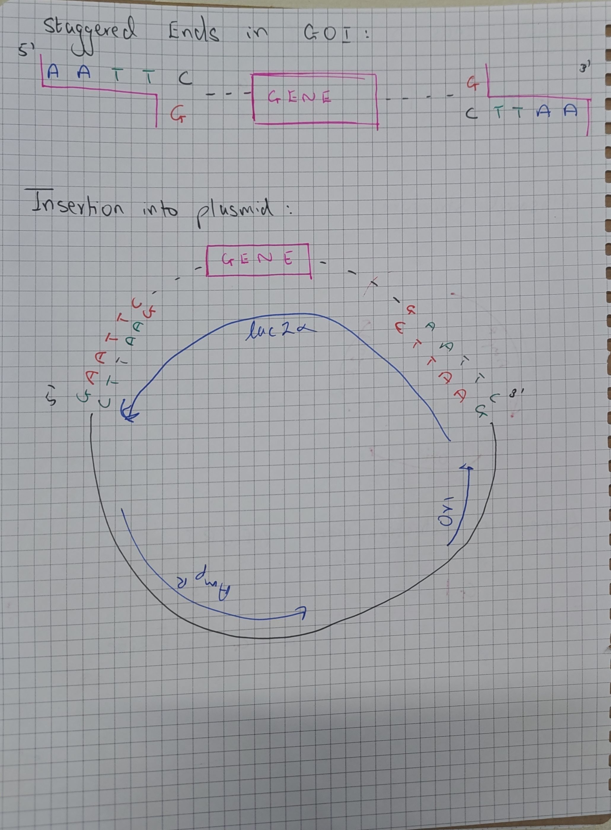
Both target DNA and pUC19 are cleaved with EcoRI, generating compatible cohesiveoverhangs. Agarose gel electrophoresis resolves fragments by molecular mass, allowing clean gel extraction of the excised insert. T4 DNA Ligase Reaction:
Purified insert and linearized vector are incubated with T4 DNA ligase in the presence of ATP. Cohesive sticky ends anneal via complementary hydrogen bonding, and ligase catalyzes covalent phosphodiester bond formation, restoring closed circular plasmids.Bacterial Transformation:
Recombinant plasmids are introduced into chemically competent E. coli DH5cells via heat-shock ( for 45s). Antibiotic Selection:
Transformed cells are plated onto solid LB media containing ampicillin (50 µg/mL). Untransformed cells lacking the-lactamase gene cannot survive. Blue-White Colony Screening (Chromogenic Selection):
Plates are supplemented with IPTG (isopropyl-D-1-thiogalactopyranoside, non-metabolizable inducer) and X-Gal (5-bromo-4-chloro-3-indolyl- -D-galactopyranoside, chromogenic substrate). - Non-recombinant recircularized plasmids: Retain functional
, producing active -galactosidase that cleaves X-Gal to produce blue colonies. - Recombinant plasmids: Insertional disruption of
prevents functional -complementation, yielding white colonies. Recombinant clones are isolated from white colonies.
- Non-recombinant recircularized plasmids: Retain functional
NOTE
Orientation Considerations:
Because a single restriction enzyme (EcoRI) is utilized for both
Schematic Transformation & Screening Flow
 |  |  |
|---|---|---|
| Ligation and recombinant plasmid generation | Heat-shock transformation into DH5-alpha | Chromogenic blue-white selection mechanism |
References
- Sambrook, J. and Russell, D.W. (2001). Molecular Cloning: A Laboratory Manual. 3rd Edition, Cold Spring Harbor Laboratory Press.
- Yanisch-Perron, C., Vieira, J. and Messing, J. (1985). Improved M13 phage cloning vectors and host strains: nucleotide sequences of the M13mp18 and pUC19 vectors. Gene, 33(1), pp. 103–119.